Breaking the Silence: Understanding Noise-Induced Hearing Loss
Joy of Hearing Team
Joy of Hearing Clinical Team
Sound serves as our primary connection to the physical and social environment, conveying language, emotion, and situational awareness. Yet, the exact mechanical energy that allows us to perceive a quiet whisper or a dynamic symphony possesses the immense capacity to inflict irreversible damage to the delicate structures of the inner ear. Noise-Induced Hearing Loss (NIHL) represents one of the most prevalent occupational and recreational health conditions globally. Unlike many forms of sensorineural hearing loss that result from genetic predispositions, systemic cardiovascular disease, ototoxic medications, or the natural aging process known as presbycusis, NIHL is uniquely characterized by its complete preventability.
Despite its preventable nature, NIHL frequently progresses in a highly insidious manner. Because the initial stages of structural damage often present without obvious physical pain, bleeding, or immediate shifts in hearing thresholds, a vast majority of individuals remain completely unaware of their deteriorating auditory health until the condition significantly impairs speech comprehension. Understanding the foundational mechanics of sound trauma, the profound physiological vulnerability of the cochlea, and the clinical strategies for prevention and intervention forms the absolute bedrock of modern audiological care.
The Intricate Anatomy and Physiology of Auditory Perception
To fully comprehend the destructive mechanisms of excessive noise, it is necessary to examine the intricate, microscopic architecture of the peripheral auditory system. When acoustic energy enters the external auditory canal, it strikes the tympanic membrane, setting the complex ossicular chain—the malleus, incus, and stapes—into rapid motion. This mechanical action serves an essential impedance-matching function; it amplifies the sound waves transferring from an air-filled medium to a fluid-filled medium, transmitting the force directly to the oval window of the cochlea, a spiral-shaped sensory organ embedded deeply within the dense temporal bone.
Within the fluid-filled chambers of the cochlea lies the highly specialized organ of Corti, the sensory epithelium responsible for converting mechanical vibrations into electrical neural signals. The organ of Corti houses two distinctly functioning types of sensory hair cells:
- Inner Hair Cells (IHCs): Functioning as the primary sensory transducers, IHCs release neurotransmitters in direct response to mechanical stimulation, sending complex auditory information along the eighth cranial nerve directly to the brainstem and ultimately the auditory cortex.
- Outer Hair Cells (OHCs): These remarkable electromotile cells act as active cochlear amplifiers. By physically expanding and contracting in synchrony with the sound wave, they sharply increase the frequency tuning and massively enhance the sensitivity of the inner hair cells to soft sounds.
Both sensory cell types feature stereocilia—microscopic, hair-like projections arranged in precise, staircase-like rows at their apical surfaces. Connected by microscopic protein filaments called tip links, these structures are exceptionally sensitive to the shearing forces generated by fluid displacement within the cochlear chambers.
The Pathophysiology of Acoustic Overexposure
When the auditory system encounters sound pressure levels exceeding clinically safe thresholds, the resulting trauma manifests through distinct, measurable physiological pathways. Cellular damage occurs either through direct mechanical destruction or profound metabolic exhaustion. The cochlea is organized tonotopically, meaning high-frequency sounds are encoded at the basal end, while low frequencies are processed at the apical end. Because all incoming sound energy must physically pass through the basal region to reach the apex, the high-frequency hair cells endure the highest amount of continuous wear and tear, making them exceptionally vulnerable to injury.
Acute Mechanical Trauma
Acute exposure to intense impulse noises—such as a gunshot, an industrial explosion, or a sudden acoustic blast from heavy machinery—can generate instantaneous, catastrophic cellular damage. At extreme sound pressure levels, typically exceeding 140 decibels (dB), the violent, uncontrolled displacement of cochlear fluids can physically rupture the fragile cell membranes. The immense pressure waves tear the stereocilia entirely from their anchoring points on the basilar membrane, breaking the essential tip links that govern ion channel activation. In the most severe instances of acoustic trauma, the entire basilar membrane can dislocate. This sudden, violent biomechanical event results in immediate, profound, and entirely permanent sensorineural hearing loss.
Chronic Metabolic Exhaustion
Conversely, chronic exposure to continuous, elevated noise levels—such as heavy machinery operation, persistently loud music, or prolonged daily use of personal listening devices—induces severe metabolic damage. The continuous sound stimulation forces the hair cells into a state of relentless hyperactivity. This extreme hypermetabolic state leads to a massive accumulation of reactive oxygen species (ROS), commonly referred to as free radicals, within the sensitive cochlear tissues.
When the local concentration of free radicals ultimately overwhelms the inner ear’s natural cellular antioxidant defense mechanisms, a state of severe oxidative stress ensues. This stress damages mitochondrial DNA, violently disrupts protein synthesis, and ultimately triggers apoptosis, the process of programmed cell death. Because human inner ear hair cells fundamentally lack the biological capacity for spontaneous regeneration, the death of these sensory receptors guarantees permanent auditory deficits.
Cochlear Synaptopathy (Hidden Hearing Loss)
Recent audiological research has identified an even more subtle, insidious form of noise-induced damage: cochlear synaptopathy. Long before the physical hair cells themselves undergo complete apoptosis, the delicate synaptic connections between the inner hair cells and the primary auditory nerve fibers can rapidly degrade and disconnect. Patients presenting with cochlear synaptopathy very frequently show seemingly normal audiometric thresholds in a quiet clinical setting but struggle profoundly to understand human speech in environments with competing background noise. The destruction of these low-spontaneous-rate neural synapses severely compromises the temporal resolution and clarity of the auditory signal reaching the brain, a phenomenon clinically classified as “hidden hearing loss.”
Clinical Manifestations and Audiometric Presentation
The progression of Noise-Induced Hearing Loss typically follows a highly predictable clinical trajectory. Initially, patients may experience what is known as a Temporary Threshold Shift (TTS). Following a specifically loud event, such as attending an amplified rock concert or completing a long shift on a noisy factory floor, an individual may acutely notice a feeling of aural fullness or a distinctly muffled quality to environmental sounds. This TTS acts as a protective physiological fatigue response, and baseline hearing sensitivity generally recovers within a 24 to 48-hour window as the delicate metabolic balance within the cochlea is slowly restored.
However, repeated instances of TTS eventually and inevitably transition into a Permanent Threshold Shift (PTS). The defining clinical hallmark of a PTS directly caused by noise exposure is a localized, sharply defined drop in hearing sensitivity, observable on a standard audiogram as the classic “noise notch.” This specific notch predominantly affects the high frequencies, typically centered directly between 3,000 Hz and 6,000 Hz, often showing a slight recovery at 8,000 Hz.
Tinnitus: The Internal Warning Signal
Tinnitus—the persistent perception of ringing, buzzing, roaring, or hissing in the ears entirely absent of any external sound source—is frequently the initial, glaring symptom of auditory distress. Tinnitus generated directly by noise exposure results from the central auditory nervous system attempting to heavily overcompensate for the sudden loss of peripheral neural input. Deprived of normal, expected sensory stimulation from the newly damaged hair cells, the central auditory cortex drastically increases its spontaneous neural firing rate, manifesting physically as an intrusive phantom sound.
Real-World Clinical Profiles
Understanding exactly how NIHL impacts daily social and occupational functioning is best illustrated through direct clinical observation. The following specific scenarios highlight the diverse populations frequently affected by acoustic trauma.
Clinical Example 1: The Occupational Hazard
John, a 52-year-old manufacturing plant shift supervisor, presented to our clinical practice reporting massively increasing frustration during daily team meetings. For well over two decades, John worked immediately adjacent to heavy steel stamping presses. Despite the general availability of protective gear on the floor, consistent usage was never strictly enforced during the early stages of his career.
Upon comprehensive audiological evaluation, John’s pure-tone audiometry predictably revealed a classic, severe bilateral sensorineural loss dipping extremely sharply at exactly 4,000 Hz. While he could effortlessly hear low-frequency vowel sounds, the high-frequency consonant markers (such as “s,” “f,” “th,” and “sh”) were severely and permanently compromised. John’s inability to differentiate these essential phonemes perfectly explained his extreme difficulty following complex conversations in the highly reverberant environment of the factory floor. The continuous, decades-long metabolic stress had permanently decimated his outer hair cells in the basal region.
Clinical Example 2: The Modern Recreational Risk
Sarah, a 24-year-old freelance graphic designer, actively sought specialized assessment for a constant, high-pitched ringing in her right ear. She regularly attended indoor, heavily amplified music festivals and routinely utilized tightly fitting in-ear monitors for eight hours daily while working, often drastically increasing the volume simply to mask ambient office chatter.
While her standard behavioral audiogram indicated thresholds strictly within the normal range, her advanced speech-in-noise testing results were markedly poor. She exhibited the classic, definitive signs of early cochlear synaptopathy. The highly prolonged, unsafe listening levels transmitted through her personal audio devices had selectively and permanently damaged the low-spontaneous-rate auditory nerve fibers, significantly degrading the clarity of complex acoustic signals without shifting her absolute volume thresholds.
Comprehensive Diagnostic Protocols in the Clinic
Accurate, definitive diagnosis and exact staging of NIHL require an extensive, multifaceted test battery. At Joy of Hearing, our specialized clinicians employ advanced diagnostic tools to actively quantify the precise nature of the auditory damage:
- Comprehensive Pure-Tone Audiometry: Conducted within a calibrated, sound-treated booth, this establishes the behavioral hearing thresholds across the entire frequency spectrum, specifically seeking the characteristic 4 kHz notch.
- Immittance Testing (Tympanometry and Acoustic Reflexes): This precisely assesses the mechanical compliance of the middle ear system to actively rule out concurrent conductive pathologies that might directly mimic or heavily exacerbate the sensorineural loss.
- Otoacoustic Emissions (OAEs): This highly objective test directly measures the microscopic, physical sounds generated by the outer hair cells in direct response to an acoustic stimulus. Absent or significantly reduced OAEs provide early, concrete evidence of microscopic cellular damage, often long before any shift is visible on the behavioral audiogram.
- Speech-in-Noise (SIN) Testing: Essential for directly detecting functional, real-world communication deficits, tests like the QuickSIN actively evaluate the central auditory system’s raw ability to process human speech amid heavily competing background noise, isolating the specific symptoms of hidden hearing loss.
Evidence-Based Preventive Strategies
Mitigating the high risk of NIHL relies entirely on the proactive management of daily sound exposure. Clinical audiologists advocate for a structured, multifaceted approach to hearing conservation based strictly on the occupational hierarchy of controls.
- Engineering and Administrative Controls: In heavy industrial settings, this involves direct physical modifications to heavy machinery to actively reduce acoustic output, constructing dense sound barriers, and implementing strictly enforced shift rotations to fundamentally limit the total daily duration of exposure for any single employee.
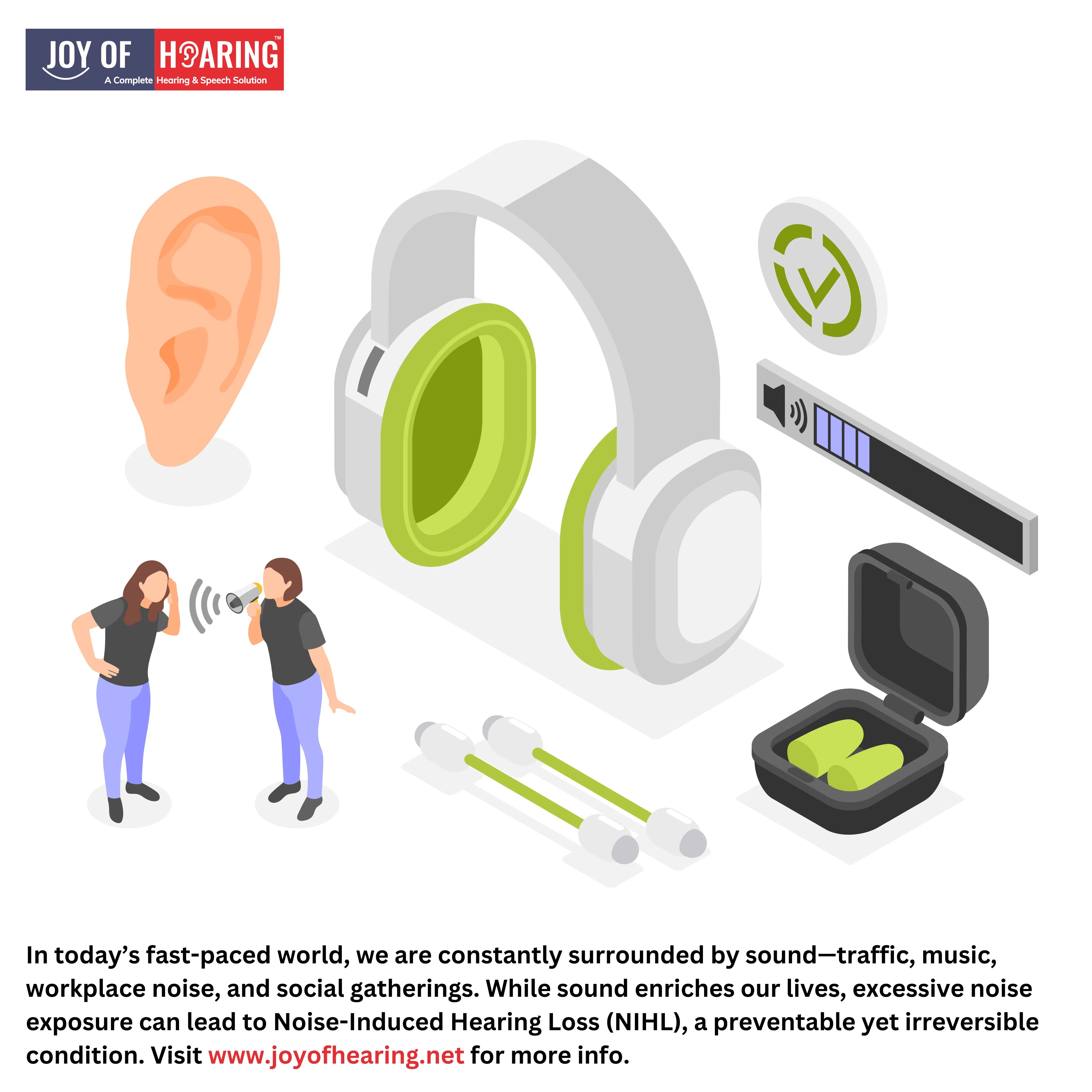
- Hearing Protection Devices (HPDs): When direct source control is functionally impossible, personal protective equipment is absolutely mandatory. High-density foam earplugs, custom-molded silicone plugs, and heavy-duty acoustic earmuffs drastically attenuate incoming sound pressure. However, HPDs must be fitted precisely by a professional to consistently achieve their designated Noise Reduction Rating (NRR).
- The 60/60 Rule for Personal Audio: For daily recreational listening, individuals must practice strict volume management. The gold-standard clinical recommendation dictates listening at strictly no more than 60% of the device’s maximum volume output for absolutely no longer than 60 minutes continuously before taking a mandated quiet break.
- Musician’s Earplugs: These highly specialized devices utilize flat-response acoustic filters to provide even attenuation. They actively reduce the overall decibel level equally across all frequencies, rigorously protecting the delicate cochlea without artificially distorting the fidelity or tone of the music.
Advanced Management and Rehabilitation
For those individuals already living with the permanent, irreversible consequences of NIHL, comprehensive, structured auditory rehabilitation is strictly required to successfully restore communication efficacy and actively prevent the severe cognitive decline heavily associated with untreated hearing loss. Untreated hearing loss imposes a massive cognitive load; the brain constantly reroutes energy to decode speech, leading to severe fatigue and a significantly increased risk of developing early-onset dementia.
Modern, highly advanced hearing aid technology utilizes sophisticated digital signal processing microchips to specifically address the physiological deficits of NIHL. Directional microphone arrays and highly aggressive noise reduction algorithms actively prioritize speech signals while simultaneously suppressing competing background noise. Frequency lowering technology can easily take entirely inaudible high-frequency consonant sounds and shift them completely into lower, much healthier frequency regions where the patient firmly retains functional, healthy hair cells.
Additionally, highly structured tinnitus retraining therapy (TRT) and customized sound masking protocols can be integrated directly into the programming of hearing instruments to provide immediate, sustained relief from continuous phantom ringing. Combined carefully with strategic auditory training exercises, motivated patients can successfully retrain their central auditory nervous system to extract meaningful information from previously degraded peripheral input.
Preserving your hearing requires conscious effort, strict daily discipline, and proactive medical care. Protecting the intricate, fragile mechanisms of the inner ear ensures that the full, vibrant spectrum of environmental and communicative sounds remains permanently accessible for an entire lifetime.